Figure 1.
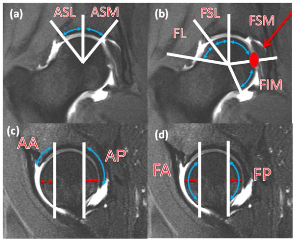
Subregions of articular cartilage. for femur on coronal (a) and sagittal (c) images. Subregions for acetabulum on coronal (b) and sagittal (d) images. (a) coronal MR image demonstrating acetabular superolateral (ASL) and superomedial (ASM) subregions divided by vertical line extending from femoral head center.(b) coronal MR image demonstrating femoral lateral (FL), superolateral (FSL) and superomedial (FSM) and inferior (FIM) subregions divided by line extending from femoral head center, to the lateral acetabular rim, to straight vertical direction and to the ligamentum teres attachment. (c) sagittal MR image demonstrates acetabular anterior (AA) and posterior (AP) subregion, demarcated by vertical line 1 cm from the most anterior and posterior aspect of the femoral head. (d) sagittal MR image demonstrates femoral anterior (FA) and posterior (FP) subregion, demarcated by vertical line 1 cm from the most anterior and posterior aspect of the femoral head.

