
Figure 5. The Extent of Singlet Oxygen-Mediated Photo-oxidation as Measured with IFP Photobleaching.
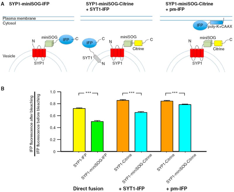
(A) Schematic drawing showing the three conditions tested in the IFP bleaching experiments. IFP was expressed in neurons either fused to the C terminus of the SYP1 or SYP1-miniSOG (left panel). In the other conditions, IFP was coexpressed as synaptotagmin-1 (SYT1) fusion (middle panel) or tethered to the plasma membrane (pm-IFP) in neurons expressing SYP1-miniSOG-Citrine or SYP1-Citrine.
(B) Summary graph showing the IFP bleaching in the six conditions tested. Significant greater IFP bleaching were observed in all three conditions. A smaller difference was observed when IFP was expressed on the plasma membrane and SYP1-miniSOG or SYP1 were expressed on the vesicles. MiniSOG was excited by 93 s of 495 nm light at 20 mW/mm2 and IFP was imaged with 665 nm excitation light.
*** indicates difference at p < 0.001. The error bars indicate SEM.