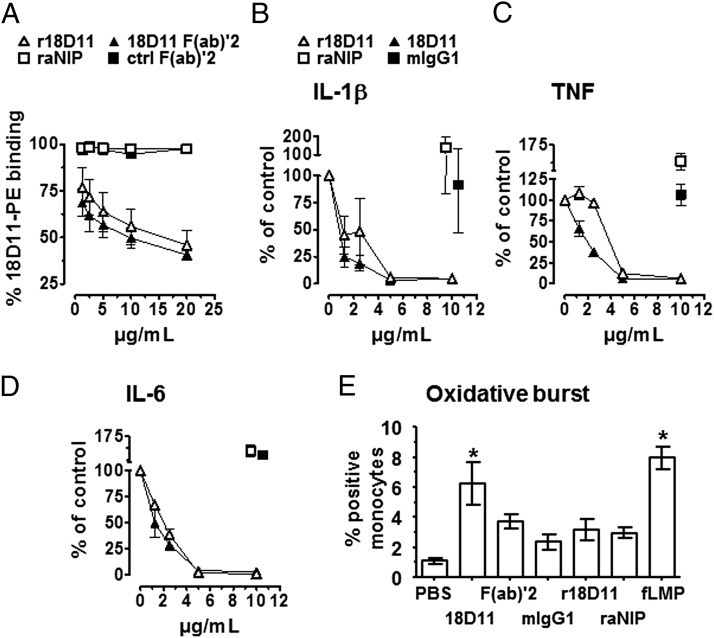
FIGURE 4.
Functional characterization of anti-human CD14 Ab r18D11. (A) Binding of increasing concentrations of r18D11 (△), raNIP (□), 18D11 F(ab)′2 (▴) or a control F(ab)′2 (▪) to monocytes was determined by the ability of the Abs to displace 10 μg/ml of the original clone 18D11 mIgG1 from its CD14 binding site in human whole blood. Mean fluorescence intensity derived from a PE-conjugated anti-mouse IgG Ab was measured using flow cytometry. Fluorescence intensity in presence of original clone 18D11 alone was set to 100%. (B–D) Release of the proinflammatory cytokines IL-1β (B), TNF (C), and IL-6 (D) from human whole blood was induced with 100 ng/ml ultrapure LPS from E. coli O111:B4 in the presence of increasing concentrations of r18D11 (△) or the original clone 18D11 (▴). raNIP (□) and mIgG1 isotype control (▪) (10 μg/ml) served as negative controls. (E) Monocyte oxidative burst was measured with flow cytometry after adding the different Ab preparations to human whole blood. Data are given as mean and SEM (n = 3 independent experiments). *p < 0.05 as compared with the negative PBS control.

