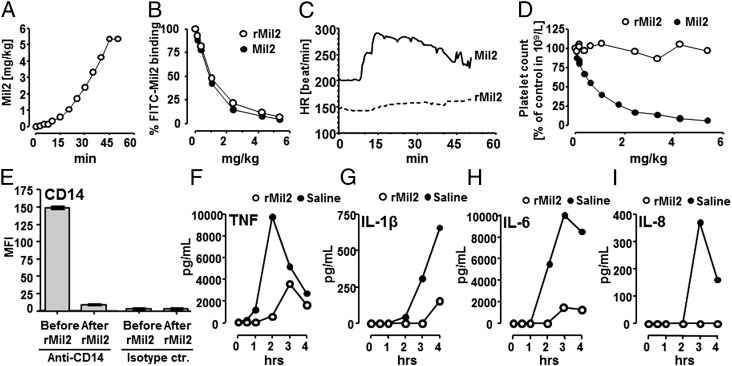
FIGURE 6.
In vivo application of anti-porcine CD14 Abs Mil2 and rMil2. Healthy newborn piglets (A–D) were infused i.v. with increasing amounts of the original clone Mil2 (n = 1) or rMil2 (n = 1) and observed for 50 min. (A) After initial small doses within the first 10 min, Mil2 or rMil2 were given every 5 min for an additional 35 min. After 45 min, a maximum final concentration of 5.36 mg/kg was reached. (B) Saturation of endogenous CD14 binding sites as a function of rMil2 (○) or Mil2 (●) concentration was measured by flow cytometry. Blood samples were incubated with 150 μg/ml FITC-conjugated Mil2, the porcine granulocyte population was selected, and the median fluorescence intensity for bound FITC-Mil2 was recorded. Fluorescence intensity in the absence of nonconjugated anti-CD14 (time point zero) was set to 100%. (C) The heart rate (HR) was recorded in real time throughout the experiments. (D) Blood platelet counts are given as a function of rMil2 (○) or Mil2 (●) concentration. They were corrected for actual hematocrit levels that reflect blood dilution caused by sampling and reconstitution. Levels in the absence of anti-CD14 Ab were set to 100%. Two piglets underwent E. coli sepsis (E–I). One piglet was injected with a bolus dose of rMil2 before i.v. infusion with bacteria and one received saline as control. Expression of leukocyte CD14 was measured before and after rMil2 injection (Fig. 6E). Cytokine release was measured during the 4-h observation period (Fig. 6F–I) comparing saline (●) with rMil2 (○). Anti-CD14, Fluorescence-labeled rMIL2; Islotype ctr, the isotype control for rMIL2.

