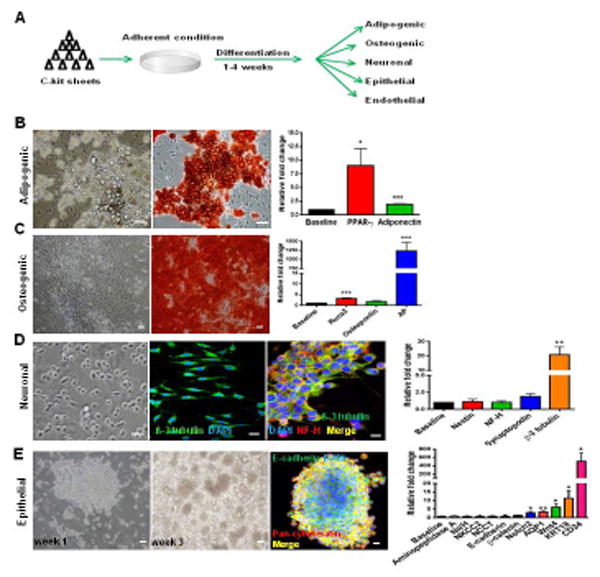
Figure 3. Non-clonal c-kit+/Lin− cells show multilineage differentiation.

(A) Schematic of the experimental procedure.
(B) Adipogenic differentiation exhibited lipid droplet accumulation, Oil Red O positivity, and PPAR-γ (*P=0.026) and adiponectin (***P<0.0001) up-regulation.
(C) Osteogenic differentiation exhibited Alizarin Red S positivity and Runx2 (***P<0.0001), osteopontin (P=0.077), and alkaline phosphatase (AP;***P<0.0001) up-regulation.
(D) Monolayer of c-kit+ /Lin- cells exhibiting prolongations in neuronal medium. β-3 tubulin was positive and co-localized with NF-H. β-3 tubulin was up-regulated (**P=0.0071).
(E) C-kit+ cells after 3 weeks in epithelial medium formed embryoid body-like structures that stained for E-cadherin and pan-cytokeratin. Notch2 (*P=0.034), AQP1 (**P=0.0056), Wnt4 (*P=0.039), KRT18 (*P=0.028), and CD24 (*P=0.035) were up-regulated.
Cell nuclei are stained blue with DAPI. Scale bars represent 50 μm (B-E) and 20 μm for confocal images (D,E). Error bars represent mean ± SEM.
