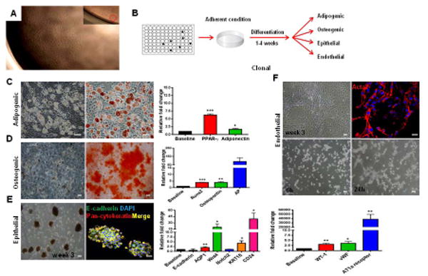
Figure 5. Clonal–derived c-kit+ cells present multipotent differentiation capacity.

(A) C-kit+ derived clones were obtained by carrying out two serial dilutions in a 96-well plate. Magnification 4x. Insert shows 1 cell/well.
(B) Schematic representation of the experimental procedure.
(C) Adipogenic differentiation exhibited lipid droplet accumulation, Oil Red O positivity, and PPAR-γ (***P<0.0001) and adiponectin (*P=0.036) up-regulation.
(D) Osteogenic differentiation demonstrated Alizarin Red S positivity, and Runx2 (***P<0.0001), osteopontin (**P=0.0012), and AP (P=0.059) up-regulation.
(E) C-kit+ clonal cells formed packed clusters that detached and acquired an embryoid body-like morphology after 3 weeks. Epithelial spheres stained for E-cadherin and pan-cytokeratin. AQP1 (**P=0.009), Wnt4 (*P=0.023), Notch2 (P=0.8), KRT18 (**P=0.0044), and CD24 (*P=0.011) were up-regulated.
(F) In endothelial medium, c-kit+ cells formed myotube-like structures that stained for Acta2. Matrigel assay revealed tube formation at 24h. WT-1 (**P=0.0036), vWF (*P=0.017), and AT1a receptor (**P=0.0065) were up-regulated.
Cell nuclei are stained blue with DAPI. Scale bars represent 50 μm for C-F and 20 μm for confocal images (E,F). Error bars represent mean ± SEM.
