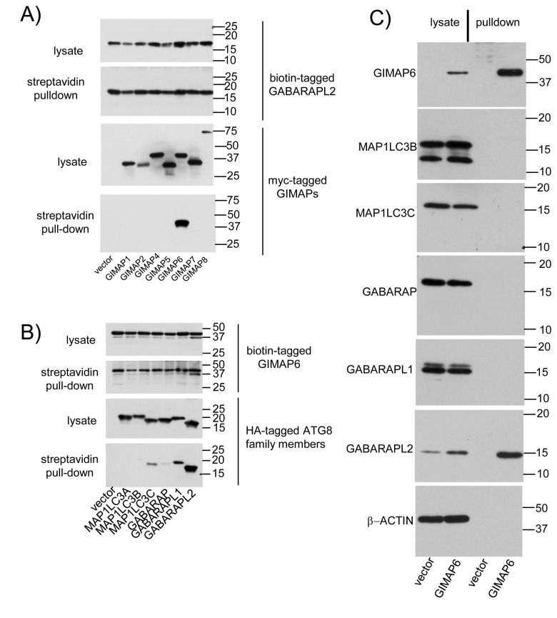
Figure 2. Specificity of GIMAP6-GABARAPL2 interactions.
A) HEK293T cells were transfected with 10 µg GABARAPL2 in pcDNA3Biot1His6iresBirA and 10 µg myc-tagged human GIMAP-encoding plasmids as indicated. 48 h later, cell lysates were prepared and biotinylated GABARAPL2 and associated proteins recovered on streptavidin-agarose beads. Aliquots of the lysates and recovered proteins were separated by SDS-PAGE and blotted for biotinylated proteins using HRP-conjugated streptavidin or myc-tagged proteins using mouse anti-myc mAb 9E10 followed by HRP-conjugated goat anti-mouse IgG. B) HEK293T cells were transfected with 10 µg human GIMAP6 in pcDNA3Biot1His6iresBirA and 10 µg HA-tagged Atg8-encoding plasmids as indicated. 48 h later, cell lysates were prepared and biotinylated GIMAP6 and associated proteins recovered on streptavidin-agarose beads. Aliquots of the lysates and recovered proteins were separated by SDS-PAGE and blotted for biotinylated proteins using HRP-conjugated streptavidin or HA-tagged proteins using mouse anti-HA mAb 12CA5 followed by HRP-conjugated goat anti-mouse IgG. Blots were developed using Immobilon ECL western blotting substrate. C) Biotinylated proteins were purified from the Biot-GIMAP6-His myc-BirA-Jurkat cell line or the corresponding vector-only cell line using streptavidin-agarose. Western blots of lysates prepared directly from the cells or of the purified proteins were then probed with HRP-conjugated streptavidin to visualise biotinylated GIMAP6 or with MAb MAC446 to GABARAPL2 or antibodies (as detailed in the Materials and Methods section) to other Atg8 family members or β-ACTIN as described by the suppliers. Results in all three panels are representative of data obtained from at least two independent experiments.

