
Fig. 2. Gustatory Neural Circuits Revealed by Genetic Tracing: Distribution of WGA in t1r3-WGA and pkd1l3-WGA Transgenic Mice.
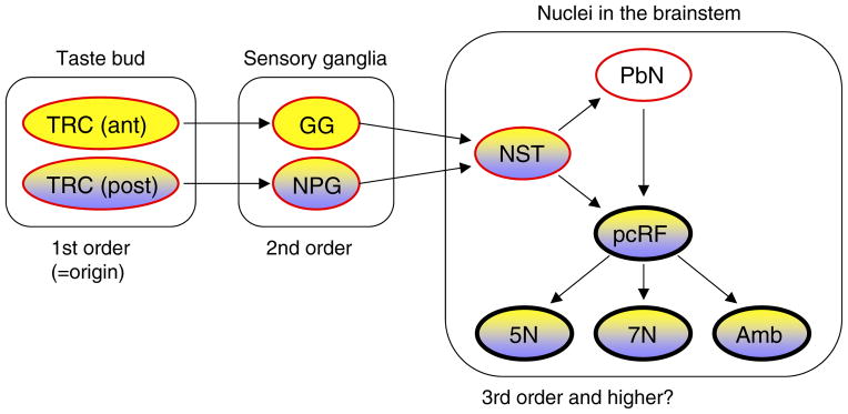
Yellow and purple indicate the tissues/nuclei where WGA immunoreactivity was detected in the t1r3-WGA and pkd1l3-WGA mice, respectively. The red outlines indicate tissues/nuclei of known ascending gustatory pathways, and arrows indicate known neural connectivity. The promoters/enhancers of t1r3 and pkd1l3 genes induced the expression of WGA mRNA faithfully in the cells where t1r3 and pkd1l3 mRNAs are endogenously expressed, and thus the WGA was observed only in taste buds of the posterior oral cavity in the pkd1l3-WGA mice. FuP, fungiform papilla; FoP, foliate papilla; CvP, circumvallate papilla; GG, geniculate ganglion; NPG, nodose/petrosal ganglia; NST, nucleus of the solitary tract; PbN, parabrachial nucleus; pcRF, parvocellular the reticular formation; 5N, trigeminal motor nucleus; 7N, facial motor nucleus; Amb, ambiguous nucleus.