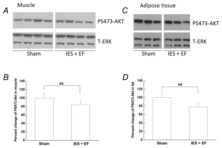
Figure 3. Insulin-induced phosphorylation of AKT was not affected by acute psychological stress in skeletal muscle and adipose tissue.
At the end of the behavioral test, 5U/kg insulin was injected and skeletal muscle and adipose tissue were removed 20 min after injection. Tissue lysates, 30 μg per lane, were subjected to Western blotting with antibodies specific for PS473-AKT or total ERK. Representative Western blots are presented in A and C. (B and D) Autoradiographs were quantified by scanning densitometry and the data are presented as the mean ± S.E of 10 mice in IES with escape failure group (IES+EF group) and 10 mice in sham group. The phosphorylated protein level in the sham group was arbitrarily set to 100%. White gap in A and C indicates grouping of lanes from different parts of the same gel.

