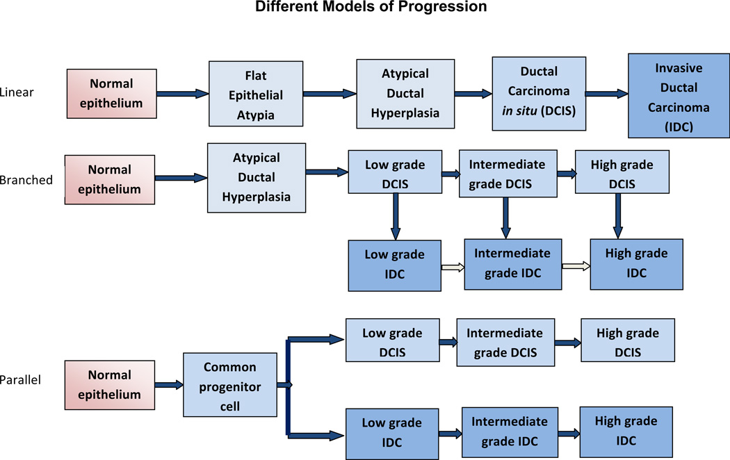
Figure 1. Models of Malignant Progression of Normal Breast Epithelium to Carcinoma.
Evidence from epidemiological, morphological and immunohistochemical studies has been used to develop several models to describe the progression from non-invasive DCIS to IDC. (Top) A linear model of progression that in sequential stages from normal epithelium to invasive carcinoma via hyperplasia and in situ carcinoma. The postulates of this hypothesis include that DCIS is a direct precursor of IDC and that atypical ductal hyperplasia is a direct precursor to low grade DCIS. In “non-linear” or “branched” models (Middle) DCIS is a progenitor of IDC, yet different grades of DCIS progress to corresponding grades of IDC. The low, intermediate and high grades can also be termed grades I, II, and III, respectively. There may also be further progression of IDC (pale arrows). The “parallel” model of progression (Bottom) hypothesizes that DCIS and IDC diverge from a common progenitor cell and progress independently through different grades in parallel.

