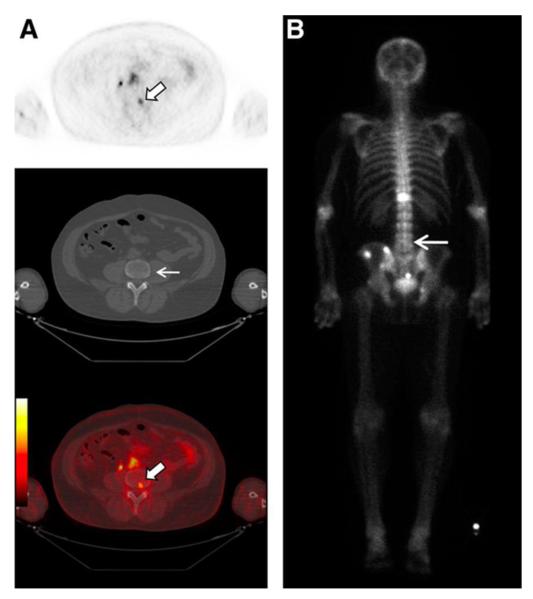
Figure 4.
18F-DCFBC PET images in a patient with progressive metastatic prostate cancer. An area of focal 18F-DCFBC uptake in the L4 vertebral body on PET and fused PET/CT (thick arrows, A) showed no correlative abnormality on CT (thin arrow, A) or bone scan (arrow, B). Reproduced with permission from Cho et al [75].

