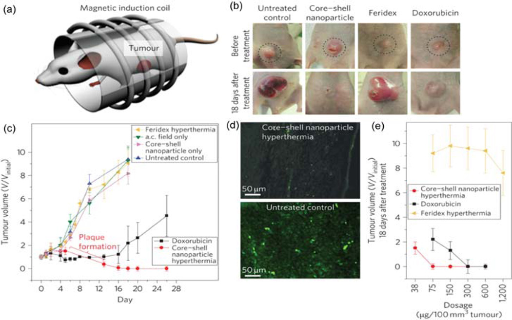
Figure 2.
(a) Magnetic hyperthermia treatment apparatus. (b) Mice xenografted with U87MG cancer cells before treatment (upper row, dotted circle) and 18 days after treatment (lower row). (c) Treatment with core–shell NP hyperthermia, Dox, Feridex hyperthermia, alternating current (a.c.) field only, core–shell NPs only or untreated control versus tumor volume (V/Vinitial). (d) Immunofluorescence images of the tumor region after nanoparticle mediated hyperthermia treatment (upper image) and the control tumor region (lower image). (e) Dose versus tumor volume measured 18 days after treatment. Reprinted with permission from [27].

