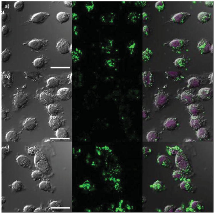
Figure 8.

Confocal microscope images of AsPC-1 cancer cells incubated with a) FITC-labeled PSQ-2, b) FITC-labeled PEG-PSQ-2, and c) FITC-labeled AA-PEG-PSQ-2. The figures from left to right depict bright field images, green fluorescence of FITC-labeled PSQ materials, and merged images of bright field and green fluorescence with DRAQ5-stained nuclei, respectively. The cells were incubated in the presence of Gd-PSQ particles for 12 h at a concentration of 100 μg/mL. Scale bar is 20 μm.
