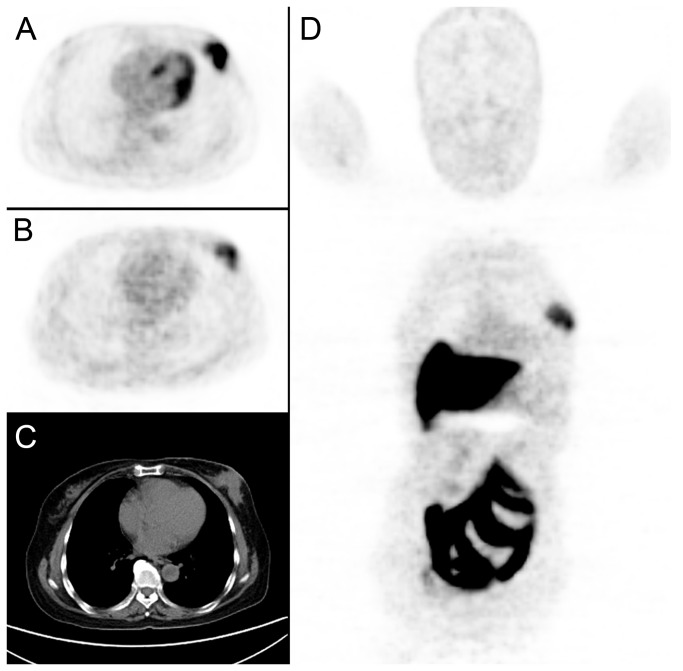
Figure 2. A 65-year-old female breast cancer patient underwent both 18F-FDG and FES PET/CT before NAC.
A tumor was detected in left breast (C. CT imaging, diameter=5.3cm), with high FDG (A. FDG imaging, SUVmax=13.51) and FES uptake (B, D. FES imaging, SUVmax=4.3). After surgery, the pathological result was confirmed to be grade C.

