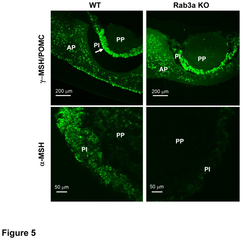
Figure 5. Alpha-MSH and pro-opiomelanocortin in Rab3a KO melanotrophs.
Representative confocal microscopy micrographs from immunocytochemistry of γ-MSH/ pro-opiomelanocortin (POMC) (top) and α-MSH (bottom) in WT and Rab3a KO pituitary slices. Arrow indicates the intermediate lobe (PI). AP=anterior part, PP=posterior part. Rab3a KO melanotrophs contained POMC, but lacked α-MSH positive signal.

