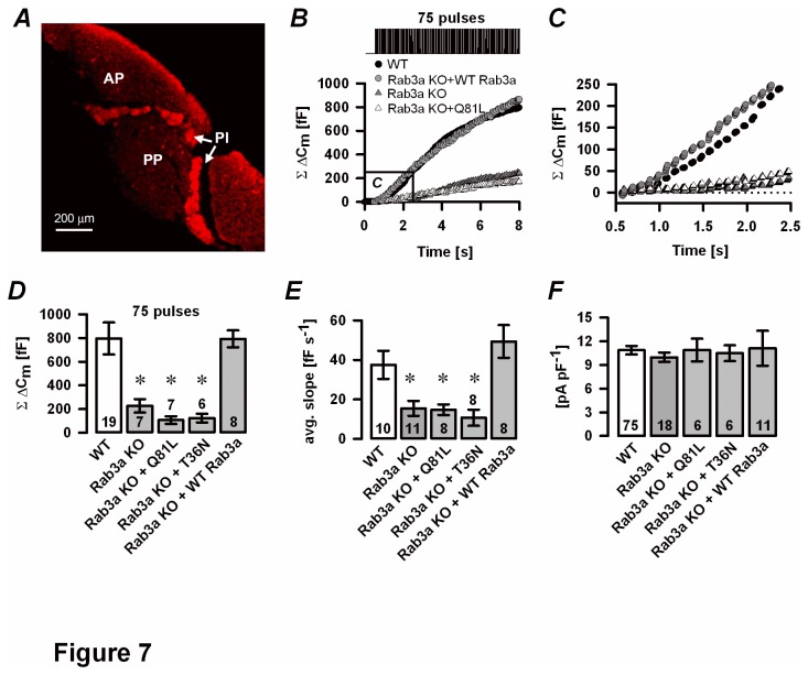
Figure 7. Rescue experiments in Rab3a KO melanotrophs.
A, Semliki forest virus transduction of a WT-Rab3a plasmid restored α-MSH in Rab3a KO pituitary slice. B, Representative cumulative ΔCm traces show depolarisation-induced secretory response in WT cells (black circles), Rab3a KO cells (grey triangles), Rab3a-deficient melanotrophs infected with Semliki forest virus harbouring a GTP-ase deficient Rab3a mutant (Rab3aQ81L, white triangles) or WT-Rab3a mutant (Rab3aKO+WT-Rab3a, grey circles). A repetitive stimulation of 75 pulses (40 ms stimulation, 10 Hz from -80 mV to 10 mV) evoked Ca2+ -dependent exocytosis. The marked area is magnified in panel C. C, The linear component was attenuated in Rab3aQ81L and Rab3a KO cells, but restored in Rab3a KO melanotrophs overexpressing a WT-Rab3a mutant. D, cumulative ΔCm. E, average slope of the linear component. F, High voltage-gated Ca2+ current density. Numbers on bars indicate the number of tested cells.∗P<0.05 versus WT.

