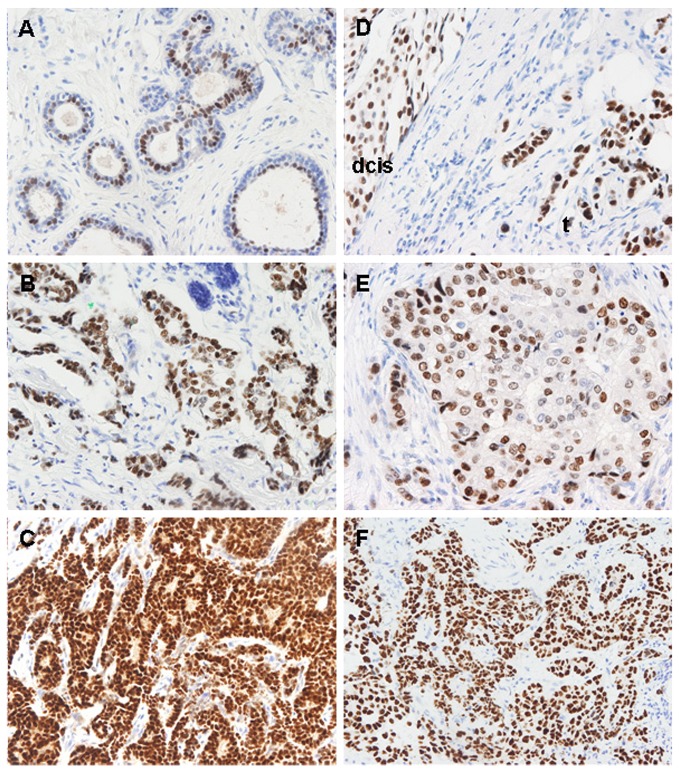
Figure 3. GATA3 immunohistochemical staining patterns in different types of breast samples and metastases.
(A) The epithelium of ducts with normal morphology shows predominant GATA3 nuclear staining in the luminal layer. (B and C). GATA3 staining in a primary tumor and its metastasis to lymph node from the same patient than A. Tumor cells are strongly positive for GATA3 both in the primary (B) and its matched lymph node metastasis (C). (D) Stronger GATA3 staining in infiltrating carcinoma (t) as compared to its associated ductal carcinoma in situ (dcis). (E) Weak GATA3 staining of a node-negative breast cancer. (F) Intense GATA3 expression in a lung metastasis of a luminal breast cancer. Most tumor cells show intense positivity.

