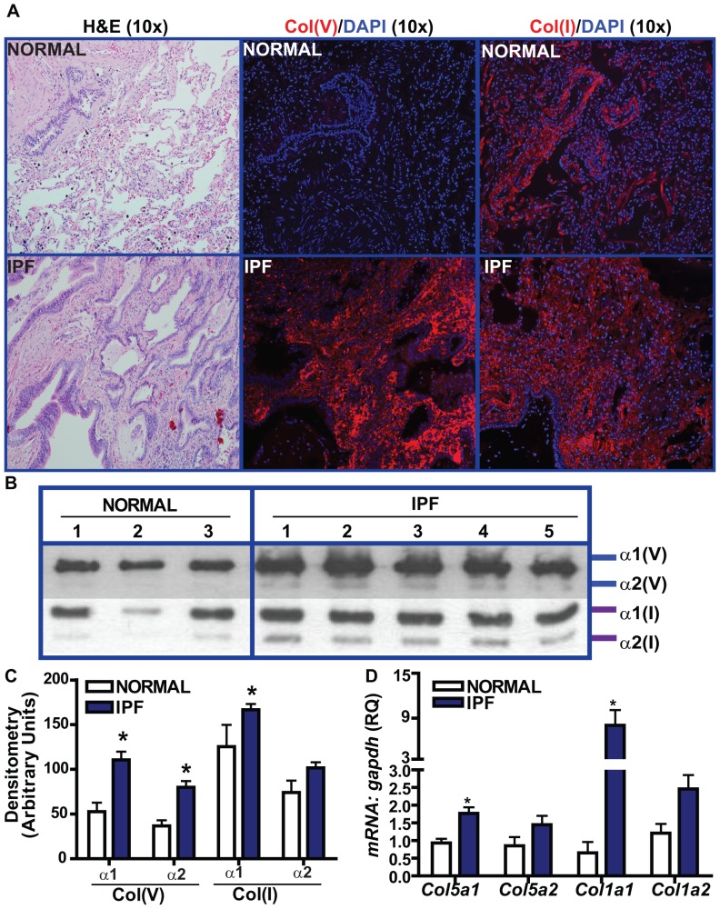
Figure 1. Relative expression of col(I) and col(V) in patients with UIP/IPF and pathologically normal specimens.
(A) Lung tissue sections from UIP/IPF patients and pathologically normal specimens were immunostained with col(I) and col(V) antibodies and their IgG, followed by incubation with rhodamine-anti-rabbit. Nuclei were counterstained with DAPI. (Original magnification, 10×, representative of 4 patients). Corresponding H&E staining is also shown. (B) Pepsin digested lung homogenates (15 µg) and corresponding standards run in a 5% gel and immunoblotted with antibodies against col(V) and col(I). Image shows representative 3 normal and 5 IPF tissues, (C) Densitometry of protein expressions of individual alpha chains of col(I) and col(V) obtained from IPF lung biopsies and pathologically normal specimens. Values represent mean ± SEM of5 normals and 20 IPF specimens (p<0.01; one-way ANOVA, post hoc test: Bonferroni), (D) mRNA expression were determined by qPCR of lung tissue sections of UIP/IPF and pathologically normal specimens. Values represent mean ± SEM; 3 normals and 4 UIP/IPF specimens; (p<0.01; one-way ANOVA, post hoc test: Bonferroni).

