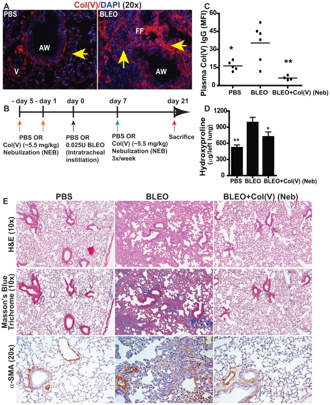
Figure 3. Tolerance induction of Col(V) protects against bleomycin-induced fibrosis.
(A) Col(V) overexpression was detected in 21 day post bleomycin injury by labeling with rhodamine and counterstained with DAPI. (Original magnification, 10×). (B) Schematic illustration of the experimental design. (C) Circulating antibodies specific to col(V) were detected in 21 day post bleomycin injury mice. Values represent mean ± SEM; number of animals: PBS = 5, BLEO = 6 and BLEO+col(V) Neb = 4. Compared to bleomycin group, * p<0.001; ** p<0.01; one-way ANOVA, post hoc test: Bonferroni. (D) Collagen deposition was measured quantitatively by assaying for hydroxyproline concentrations from the whole left lung day 21 post bleomycin injury+col(V) treatment. Values represent mean ± SEM; n = 7 mice/group. Compared to bleomycin group, ** p<0.01; * p<0.05 one-way ANOVA, post hoc test: Newman-Keuhl's. (E) At day 21 post bleomycin injury, lung tissue sections were stained for H&E and Masson's blue trichrome staining. Extensive collagen deposition was observed with bleomycin injury while col(V) nebulized lungs, similar to normal lungs, had collagen deposition around airways and vasculature. Original magnifications: 10×. (Figure S2A: 1×). Lung tissue sections were immunostained against alpha-smooth muscle actin (α-SMA) or IgG. Streptavidin-conjugated horseradish peroxidase was used with 3,3′-diaminobenzidene as substrate (brown) and nuclei were counterstained with hematoxylin (blue). Original magnifications: 20×.

