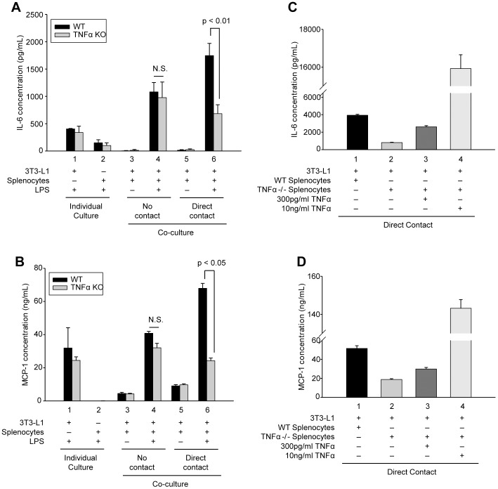
Figure 4. Cell contact-mediated enhancement of IL-6 and MCP-1 secretion requires TNFα signaling.
(A and B) Differentiated adipocytes or murine splenocytes (black bars, isolated from wild type mice; gray bars, isolated from TNFα -/- mice) were cultured alone (individual culture, columns 1 and 2) or in co-culture with no contact (columns 3 and 4) or direct contact (columns 5 and 6) as in Figure 1. Cells were incubated in the absence (−) or presence (+) of LPS (1 µg/mL) for 24 h as indicated. (C and D) Wild type (WT) or TNFα -/- (TNFα KO) splenocytes were incubated with adipocytes with direct contact in the presence of LPS (1 µg/mL) and co-cultures were supplemented with 0, 300 pg/mL or 10 ng/mL purified murine TNFα as indicated. Secreted IL-6 (A and C) and MCP-1 (B and D) were quantified by capture ELISA. Experimental points were measured in triplicate (A and B) or duplicate (C and D) for calculation of means and standard deviations. For (A and B) comparison between individual cultures, co-cultures with no contact and co-cultures with direct cell-cell contact from WT and TNFα KO were calculated using ANOVA and followed by the post-hoc Bonferroni test. For C and D all experimental points were compared statistically by ANOVA followed by the Bonferroni post-hoc test, and differed significantly (p<0.01). N.S. denotes no statistical difference.

