
Figure 1. Expression of BD-1 in the uninfected urinary tract.
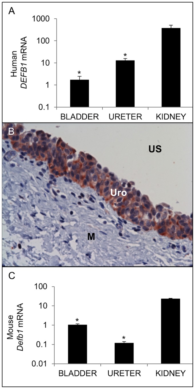
(A) Expression of human DEFB1 mRNA (TOP) and mouse Defb1 mRNA (BOTTOM). Samples were normalized for GAPDH / Gapdh content and expressed as fold-difference compared to a pool of uninfected human / mouse bladder cDNA using the 2^-ΔΔCT method[22]. * indicates p < 0.05 in 2-tailed student’s t-tests comparing indicated organ to kidney. The average fold change ± standard error of the mean (S.E.M.) for each organ is shown (n=4 bladders, 2 ureters, 3 kidneys). (B) HBD-1 protein localizes to bladder urothelium by IHC. US: Urinary Space; Uro: Urothelium; M: Muscularis. Similar results were seen in ureter (data not shown). 400x original magnification.