
Fig. 2.
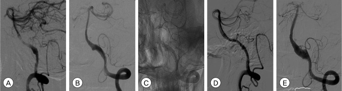
(A, B) Initial angiograms show bilateral intracranial vertebral artery (VA) dissection. The risk of rupture was higher for the left VA because the size of the aneurysm was larger on the left side. Nevertheless, we were not certain about which side had experienced rupture (A: right, B: left). (C): Native image after bilateral VA treatment shows that 2 self-expanding nitinol stents (SESs) on the left VA and 2 balloon-mounted coronary stents (BMSs) on the right VA were deployed across the dissecting lesion. (D, E) Two-month follow-up angiograms show decreased but notable contrast filling to the aneurysm sac through the stent and increased aneurysm sac filling (D:right, E:left).