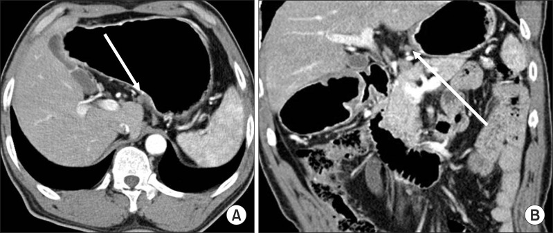
Fig. 3.
Clinical N staging using stomach protocol computed tomography: Contrast-enhanced computed tomography shows a 5.3 mm sized lymph node in infrapyloric area (arrows in A, B). The computed tomography number of this lymph node was measured about 115 HU on the average. This was revealed metastatic lymph node by pathological examination.

