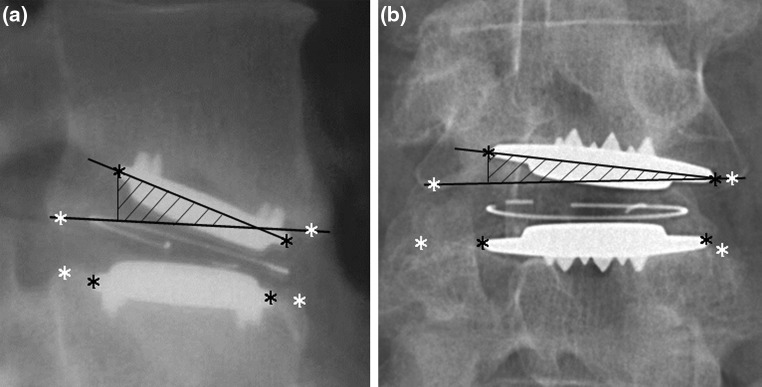
Fig. 2.

a Angle between endplate and bony endplate (α lateral) on lateral radiograph. The shading on a and b represents the calculated penetrated bone volume (PBV).The penetrated part of the TDR endplate was perpendicularly projected on the plane representing the bony endplate and the penetrated volume was calculated by integration of the orthogonal distance between the TDR endplate and this projection over the projected endplate area. b Rotation around the x-axis is measured on AP radiograph (α AP). The bony endplate of the vertebra was defined as the most superior-anterior point till the most superior-posterior point
