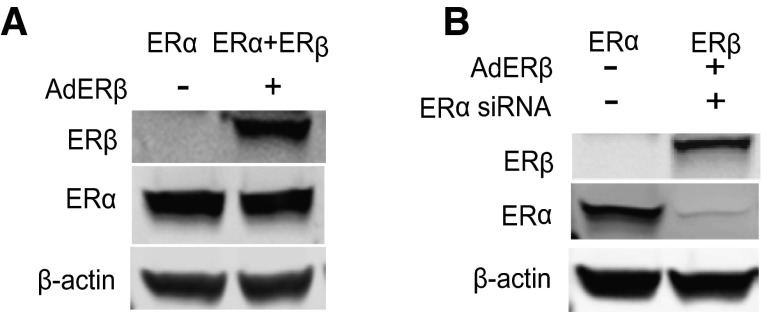
Figure 3.
Western blots show ERα and ERβ levels in cells with the 3 complements of ERs. A) MCF-7 cells were infected with control adenovirus or ERβ-expressing adenovirus to generate cells containing ERα only and ERα + ERβ, respectively. B) Cells designated as ERβ only were generated by knockdown of ERα using ERα siRNA and then infection with ERβ adenovirus.

