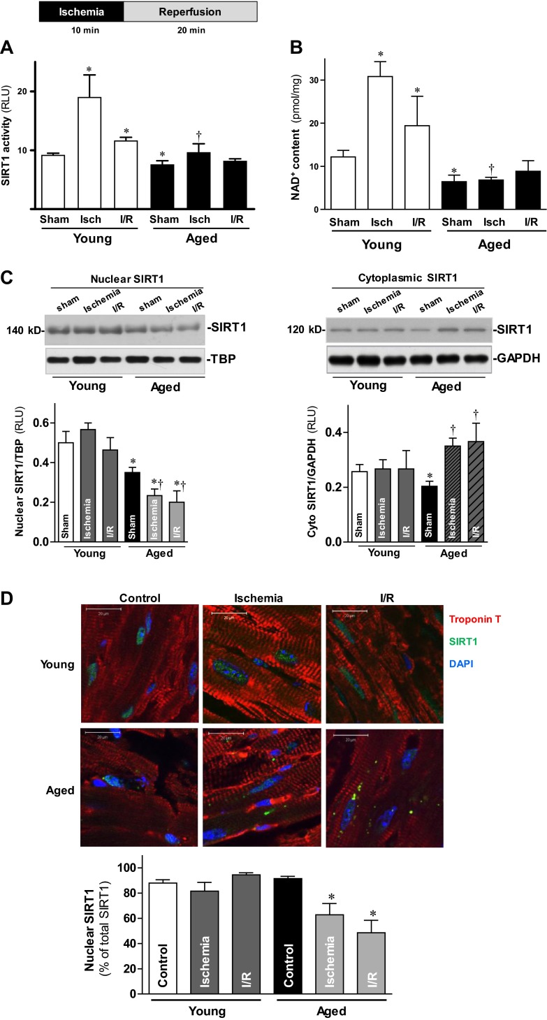
Figure 3.
Activation and nucleocytoplasmic shuttling of SIRT1 during I/R. A) LVs from young and aged hearts were harvested following a sham operation (sham), after 10 min in vivo regional ischemia (isch), and after 10 min of in vivo regional ischemia followed by 20 min reperfusion (I/R). Nuclear fractions of young and aged LVs were subjected to the SIRT1 activity assay. B) Intracellular NAD+ content measurement of LVs from young and aged heart samples were obtained from the same three aforementioned conditions. Values are means ± se; n = 6. *P < 0.05 vs. young sham; †P < 0.05 vs. young ischemia. C) Nuclear (left panel) and cytoplasmic (right panel) extracts from young and aged hearts were analyzed by immunoblotting; bar graphs show relative levels of nuclear SIRT1 (left panel) and cytoplasmic SIRT1 (right panel) from young and aged hearts. Values are means ± se; n = 6–8. *P < 0.05 vs. corresponding young group; †P < 0.05 vs. aged sham. D) Hearts from young and aged C57BL/6 mice were subjected to ex vivo global I/R with a Langendorff heart perfusion system. Hearts from control, ischemia (20 min), and I/R (20 min of ischemia followed by 30 min of reperfusion) treatment groups were collected for SIRT1 immunostaining (top panel); bar graph shows percentage of nuclear SIRT1 as a fraction of the total SIRT1 (bottom panel). Values are means ± se for 5 independent experiments. *P < 0.05 vs. aged control.

