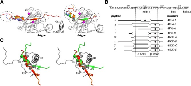
Figure 3.
Peptides bound to MMP-13 are prodomain derived. A) Cartoon representation of the peptides in A- and B-type molecules. MMP-13(E223A) is shown as a gray cartoon with Zn2+ as purple spheres. A-type molecules: peptide a (red), peptide c (green), peptide w (orange), and peptide y (blue). B-type molecules: peptide b (red), peptide d (green), peptide x (orange); peptide z (blue). Dashed lines: parts of the peptides that contribute to crystal packing. B) Schematic of the active site peptides. Sequence shows the MMP-13 prodomain with predicted helix 1. The 8 peptides are shown schematically, with the S1′-bound residue in bold and as a solid circle. C) Superimposed prodomains of MMP-2 (dark gray) showing helices 1–3 (H1–H3) in walleye stereo. The zymogen-maintaining cysteine (Cys) is indicated. Representative A- and B-type peptides are superimposed on H1. Active site cleft binding β-strand section with the P1′ Tyr residue is indicated; arrows indicate cleavage sites.

