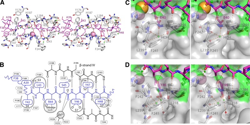
Figure 4.
Peptide binding the MMP-13 active site cleft subsites. A) Peptide d as a representative example (purple sticks and labels), Cat domain residues with at least 1 atom within 4 Å (gray sticks and labels), and hydrogen bonds between the peptide substrate and MMP-13(E223A) Cat domain (broken orange sticks). B) Schematic representation of the interactions shown in A. C) Stereo close-up of the S1′ pocket with E40 of peptide a. Water (small green spheres) and MMP-13 surface within 4 Å of the peptide (green shading). D) S1′ pocket with Y46 bound in stereo (peptide d).

