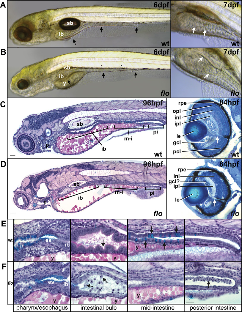
Figure 1. Intestinal abnormalities and microphthalmia in flo embryos.
DIC images of wildtype and flo embryos at 6–7dpf. (A, B) The black arrows indicate, from L→R, the three broad regions of the intestine: the intestinal bulb, mid-intestine and posterior intestine. (A) The intestinal epithelium in wildtype is extensively folded (white arrows) and is thinner and unfolded in flo (white arrows). (B) In flo, yolk resorption is incomplete and the swim bladder rarely inflates. Microphthalmia is evident and the head is slightly smaller and misshapen. (C, D left panels) Sagittal histological sections of wild-type and flo embryos at 96hpf stained with alcian blue periodic acid Schiff reagent showing the entire intestinal tract. (C, D right panels) Transverse sections of wildtype and flo eyes stained with methylene blue/azureII show disrupted cell layers in flo. White arrow indicates optic nerve. (E, F) The wildtype intestinal bulb epithelium is elaborating folds (arrow) at 96hpf, but is thin and unfolded in flo. Fewer goblet cells (turquoise staining, arrows) are present in the pharynx and mid-intestine (F) compared to wildtype (E). Detached cells in the intestinal lumen in flo (F, arrows) are not observed in wildtype (E). (es) esophagus; (gcl) ganglion cell layer; (ipl) inner plexiform layer; (inl) inner nuclear layer; (ib) intestinal bulb; (le) lens; (opl) outer plexiform layer; (pcl) photoreceptor cell layer; (p) pharynx; (rpe) retinal pigmented epithelium; (sb) swim bladder; (y) yolk. Scale bars: C, D 10µm; E, F 20µm (shown in F, last panel).

