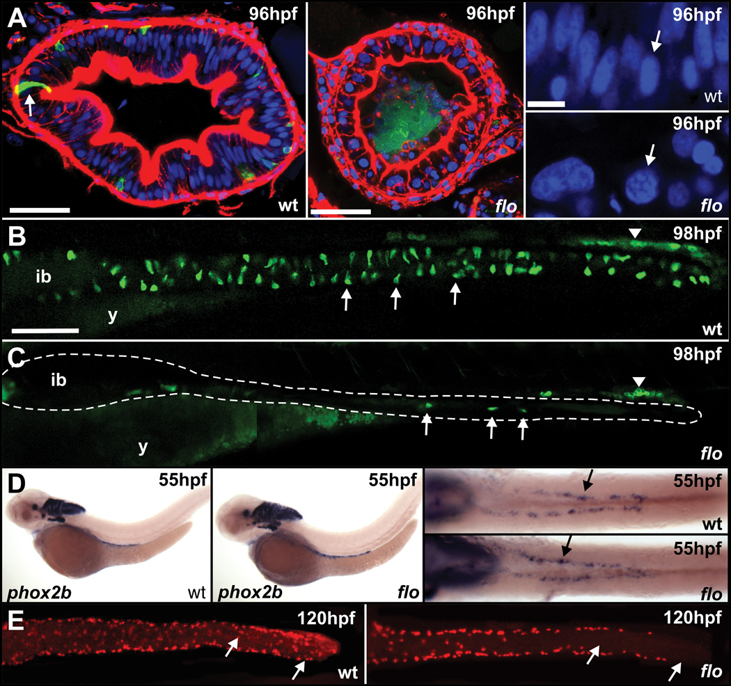
Figure 2. Abnormal cell polarization, nuclear morphology and differentiation in flo intestinal epithelium.
(A) Transverse sections through the intestinal bulb of flo;Tg(nkx2.2a:mEGFP) embryos at 96hpf, stained with rhodamine-phalloidin (red) to visualize F-actin and Hoechst 33342 (blue) to visualize DNA. Compared to wildtype, cellular organization of the intestinal epithelium in flo is disrupted. White arrow in wildtype indicates an Nkx2.2a-mGFP-positive enteroendocrine cell; in flo the GFP fluorescence is associated with cellular debris in the lumen. (A, right panels) Higher magnification images show nuclei (white arrows) are misshapen and of more variable size in flo compared to wildtype. (B, C) Fewer nkx2.2a-mEGFP-enteroendocrine cells are present in the intestine of flo;Tg(nkx2.2a:mEGFP) embryos (dashed line) compared to wildtype. nkx2.2a-mEGFP positive cells associated with pronephric ducts (white arrowheads) are unaffected in flo. (D) Lateral and ventral views of the vagal region of 55hpf embryos showing similar distribution of phox2b-expressing cells in flo and wildtype as they migrate along the intestine. Ventral views of dissected intestines from 120hpf wildtype (E, left panel) and flo (E, right panel) stained with anti-Hu antibody, showing a reduced and abnormal distribution of differentiated enteric neurons (ENS) in the mid and posterior intestine of flo embryos at 120hpf (white arrows). The numbers of ENS in a 10-somite mid-intestinal segment are: wildtype = 139 ± 3 (mean ± S.D); flo = 93 ± 5 (n = 4; Student’s t-test P<0.001). In a 10-somite segment in the posterior intestine, the numbers are: wildtype = 74 ± 8; flo = 39 ± 7 (n = 4; Student’s t-test P<0.01).

