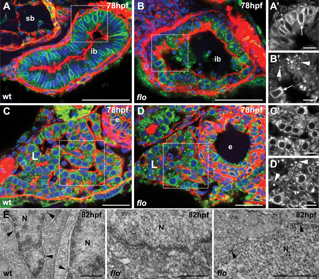
Figure 5. Aberrant distribution of nuclear pore complexes in the intestinal epithelium and liver of flo embryos at 78hpf.
(A–D) Thick transverse sections of wildtype and flo embryos stained with rhodamine-phalloidin to detect F-actin (red), Hoechst 33342 to detect DNA (blue) and the monoclonal antibody, MAb414, to detect nuclear pore complex (NPC) proteins (green). Sections (200 µm) in the region of the intestinal bulb reveal a punctate rim of fluorescence around the nuclei of cells (arrow) in wildtype intestinal epithelium (A, A’ [boxed area in A]) and liver (C, C’) but severe disruption of this pattern in the intestinal epithelium (B, B’) and liver (D, D’) of flo embryos with staining largely associated with large cytoplasmic aggregates (arrowheads) and cellular debris. (E) TEM reveals abundant nuclear pores (arrowheads) in ultrathin sections of intestinal epithelial cells in the region of the intestinal bulb of wildtype embryos but none in the corresponding tissue in flo. Meanwhile, nuclear pores are evident in a periderm cell in flo (E, right panel). Scale bars A, B = 50 µm; C, D = 25 µm; A’–D’ = 10 µm; E (left panel) = 2 µm; E (centre and right) = 0.5 µm. (N) nucleus; (ib) intestinal bulb; (L) liver; (e) esophagus; (sb) swim bladder.

