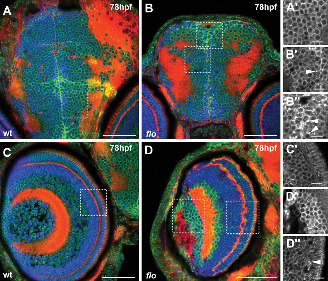
Figure 6. Aberrant distribution of nuclear pore complexes in discrete regions of the brain and eye of flo embryos at 78hpf.
Transverse sections of wildtype and flo embryos were analyzed as described in the legend to Figure 6. (A, A’) NPC localization in the wildtype midbrain produces a punctate rim of fluorescence around the nuclei of cells. A wildtype distribution is also seen in ventral areas of the flo midbrain (B, lower inset B’) but in neuroproliferative zones (dorsal midbrain and midline between the two hemispheres), cytoplasmic aggregates are found (B, upper inset B’’, arrowheads). (C, D) Similarly in the eye, some areas of wildtype NPC localization are seen in flo embryos (D, left inset D’) but in the photoreceptor cell layer (D, right inset D’’) cytoplasmic aggregates predominate. Scale bars A–D = 50 µm, A’–D’’ = 10 µm.

