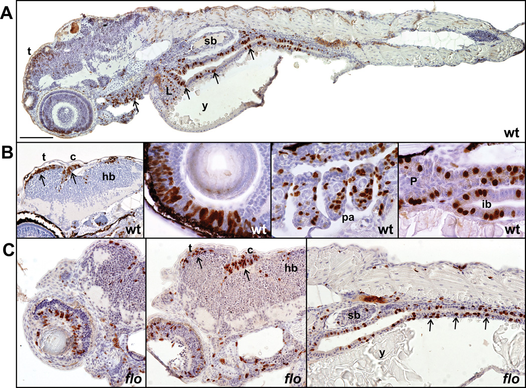
Figure 7. Localized proliferative activity in wildtype and flo embryos at 72hpf.
Sagittal sections of wildtype and flo zebrafish embryos showing BrdU-positive nuclei (brown) in cells captured in the S-phase of the cell cycle. (A–B) A high proportion of proliferative cells is observed in the entire intestinal epithelium (A, arrows; B, fourth panel), dorsal midbrain (tectum), cerebellum, dorsal hindbrain (B, first panel), retinal epithelium (B, second panel), pharyngeal arches (A, left arrow; B, third panel), liver (A), pancreas (B, fourth panel). (C) The same tissues are proliferative in flo at 72hpf. (c) cerebellum; (hb) hindbrain; (ib) intestinal bulb, (L) liver; (P) pancreas; (pa) pharyngeal arches; (sb) swim bladder; (t) tectum; (y) yolk.

