Figure 1.

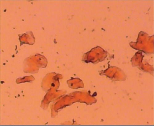
Photomicrograph of oral buccal cells from Toombak user dipping area showing the keratinization. Anucleated cells appeared in the field (Pap stain 100x).

Photomicrograph of oral buccal cells from Toombak user dipping area showing the keratinization. Anucleated cells appeared in the field (Pap stain 100x).