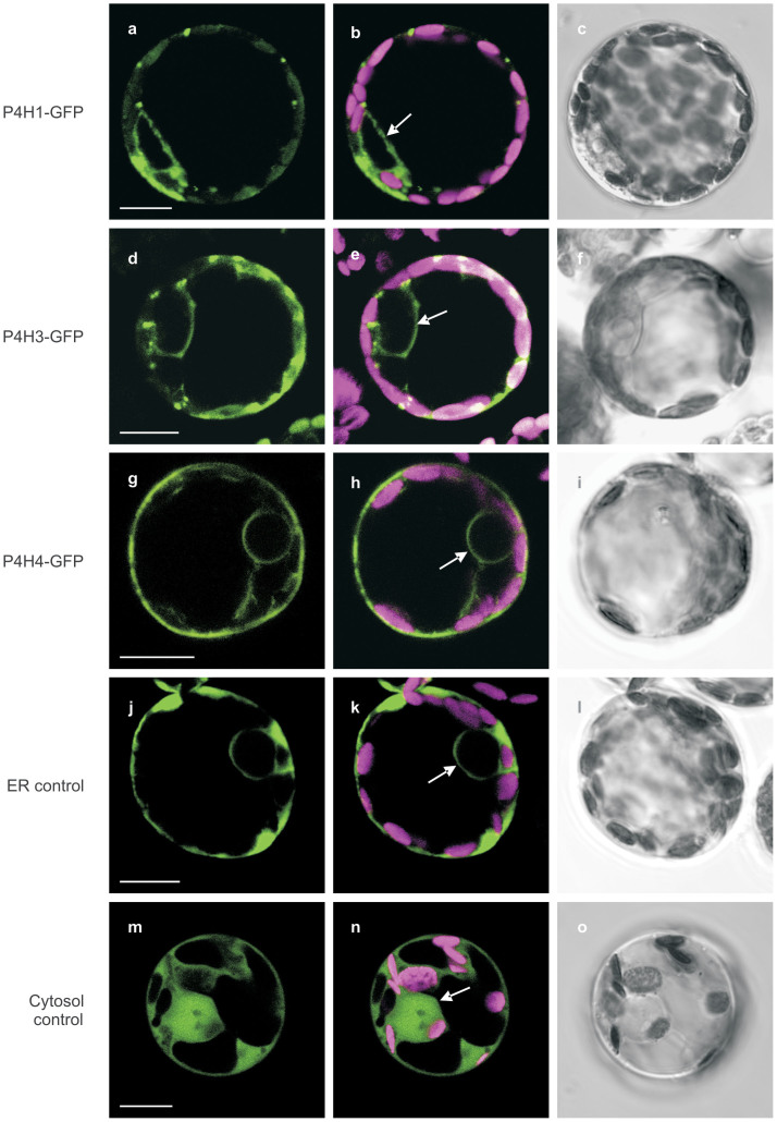
Figure 1. Subcellular localization of P. patens P4H homologues.
Fluorescence of P4H-GFP fusion proteins in P. patens protoplasts was observed by confocal microscopy 3 to 14 days after transfection. The images obtained for P4H1-GFP, P4H3-GFP and P4H4-GFP are taken as example of the fluorescence pattern which was observed for all homologues. (a–c) P4H1-GFP, (d–f) P4H3-GFP, (g–i) P4H4-GFP, (j–l) ASP-GFP-KDEL as control for ER localization, (m–o) GFP without any signal peptide as control for cytosolic localization. (a, d, g, j and m) single optical sections emitting GFP fluorescence (494–558 nm), (b, e, h, k and n) merge of chlorophyll autofluorescence (601–719 nm) and GFP fluorescence, (c, f, i, l and o) transmitted light images. The arrows indicate the cell nucleus membrane.

