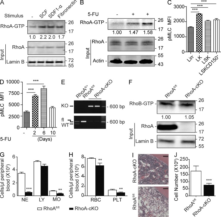
Figure 1.
RhoA deficiency causes acute hematopoietic failure. (A) Isolated Lin− cells were stimulated with SCF, SDF-1α, and fibronectin, and relative RhoA activity was determined by normalizing to total RhoA input. (B) Low-density monocytes were isolated 6 d after 5-FU injection, and relative RhoA activity was normalized to total RhoA input. (C) Phosphorylation of MLC (Ser19) in the WT primitive HSPC populations was determined by flow cytometry. MFI, mean fluorescence intensity. (D) Kinetics of MLC phosphorylation (Ser19) in Lin−SLAM population after 5-FU treatment was determined by flow cytometry. (E) RhoA deletion efficiency of the Mx-cre+RhoAfl/fl mice was determined by genotyping PCR using Lin− cells 3 d after induction. (F) RhoB activity was assessed using Lin− cells isolated 3 d after poly I:C induction. RhoB activity was determined and normalized against Lamin B. (G and H) PB counts of the congenic transplantation recipients. CD45.2+ RhoAfl/fl; Mx-cre+ or Mx-cre− cells were transplanted into lethally irradiated CD45.1+ WT recipients. Three poly I:C injections were administrated 2 mo after transplantation. Recipients were sacrificed for analysis at 5 d after the last poly I:C injections. NE, neutrophils; LY, lymphocytes; MO, monocytes; RBC, red blood cells; PLT, platelets. (I) Representative H&E staining of femur sections. Bars, 40 µm. (J) Absolute number of BM white cells in the tibia, femur, and iliac crest 5 d after three poly I:C injections. Numbers of samples analyzed: four (C and D) or five (G, H, and J) per group. (A–D, F–H, and J) The results from a representative experiment of two independent experiments are shown. (A, B, and F) Molecular masses (kilodaltons) are indicated to the right of the blots. Error bars indicate SEM. **, P < 0.01; ***, P < 0.001.

