Figure 1.
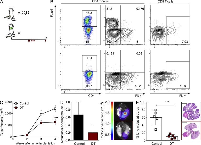
Ablation of T reg cells affects the growth of fully established primary and lung metastatic tumors. (A) Schematic of the experimental set up. Black arrows indicate day of tumor implantation (↓) and analysis (↑). Red arrows indicate regimen of DT treatment (days 14, 15, 17, 19, and 25 after tumor cell implantation). (B) Flow cytometric quantification of intratumoral CD4+ Foxp3+ T cells (left) and IFN-γ production in T cells (right). Top: control; bottom: DT-treated. (C) Growth kinetics of orthotopic tumors in mice treated with 50 µg/kg DT when tumors reached a volume of ∼250 mm3. A representative of two independent experiments is shown; n = 5 mice per group; ****, P < 0.0001. Error bars represent SEM. (D) Fraction and representative image of mice with detectable lung metastasis upon bioluminescence imaging of the dissected lungs from the group depicted in C. Error bars represent SEM. (E) Histological quantification and representative H&E staining image of the area of the lungs occupied with tumors in experimental lung colonization experiments; bars, 5,000 µm; ***, P < 0.001. Error bars represent SD. Mice were injected with 5 × 10⁵ PyMT-derived cancer cells in the tail vein and treated with DT 2 wk after tumor injection with the schedule shown in A. P-values were calculated using ANOVA, followed by Bonferroni’s post-hoc test (C) and Student’s t test (E).

