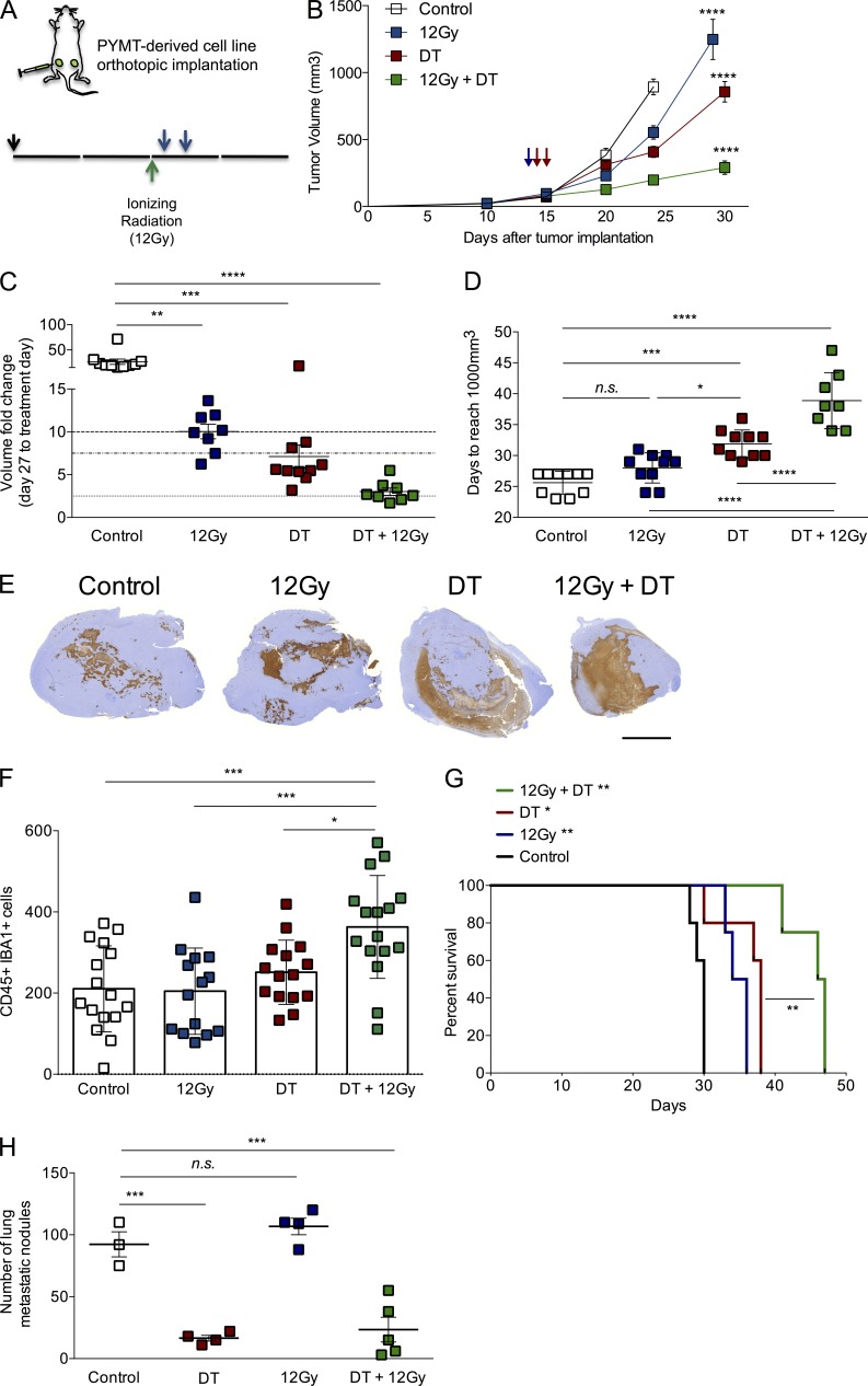
Figure 7.
T reg cell ablation improves the effect of ionizing radiation on tumor growth. (A) Scheme of treatment. 12Gy of ionizing radiation was delivered in a single fraction when tumors reached a volume of ∼100 mm3 (green arrow), followed by two i.v. injections of 25 µg/kg DT (blue arrows). Orthotopic tumor transplantation is indicated by a black arrow (↓). (B) Tumor growth kinetics in mice subjected to radiation alone, DT-mediated T reg cell ablation alone, a combination of both, or no treatment. ****, P < 0.0001. (C) Analysis of fold change increase in tumor size for each group at day 27 after initial treatment. **, P < 0.01; ***, P < 0.001; ****, P < 0.0001. (D) Day at which a given tumor reaches a volume of at least 1,000 mm3. n.s., not significant; *, P < 0.05; ***, P < 0.001; ****, P < 0.0001. (E) Representative images of histological staining with cleaved Caspase 3, depicting the area of apoptotic cells observed in each individual tumor. n = 3–5 mice per group. Bar, 5,000 µm. (F) Histological assessment of the number of CD45+ IBA1+ cells in representative viable regions of the tumor. *, P < 0.05; ***, P < 0.001. (G) Survival analysis of mice in each of the animal groups described above. *, P < 0.05; **, P < 0.01. (H) Time-matched quantification of metastatic nodules in lungs from mice in each group. ***, P < 0.001. A representative of two independent experiments is shown; n = 5 mice per group. Error bars represent SEM. P-values were calculated using ANOVA followed by Bonferroni’s post-hoc test.

