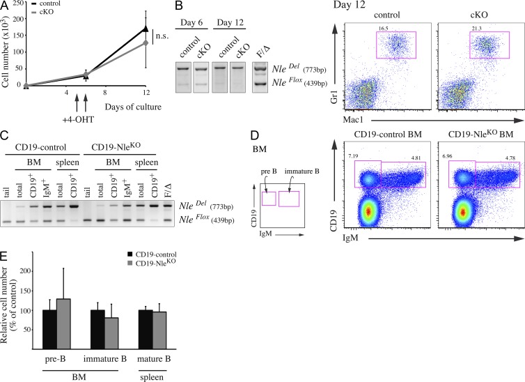
Figure 5.
Nle is dispensable during myeloid and B cell differentiation. (A and B) 200 sorted LSK Flt3− cells per well were plated onto OP9 cells. After 5 d, 4-OHT was added to the culture medium for 2 d. (A) Proliferation curves (n = 4–9 from two independent experiments). SD is shown. n.s., not significant; P = 0.211. (B) Deletion was evaluated by PCR and myeloid differentiation was assessed at day 12 by flow cytometry after staining with Mac1 and Gr1 markers. Similar results were obtained in two independent experiments. (C–E) Analysis of CD19-control (CD19Cre/+; NleFlox/+) and CD19-NleKO (CD19Cre/+; NleFlox/null) mice. (C) PCR analysis on IgM+ and CD19+ sorted cells from BM and spleen. Data are representative of three individuals. (D) Representative FACS profile of CD19-control and CD19-NleKO BM, stained with IgM and CD19 markers. (E) Counts of pre-B cells (CD19+ IgM−), immature B cells (CD19+ IgM+) from BM, and mature B cells (CD19+ IgM+) from spleen of CD19-control and CD19-NleKO mice. Bars are means (SD) of n = 4–6 mice per genotype pooled from three independent experiments.

