Figure 1.
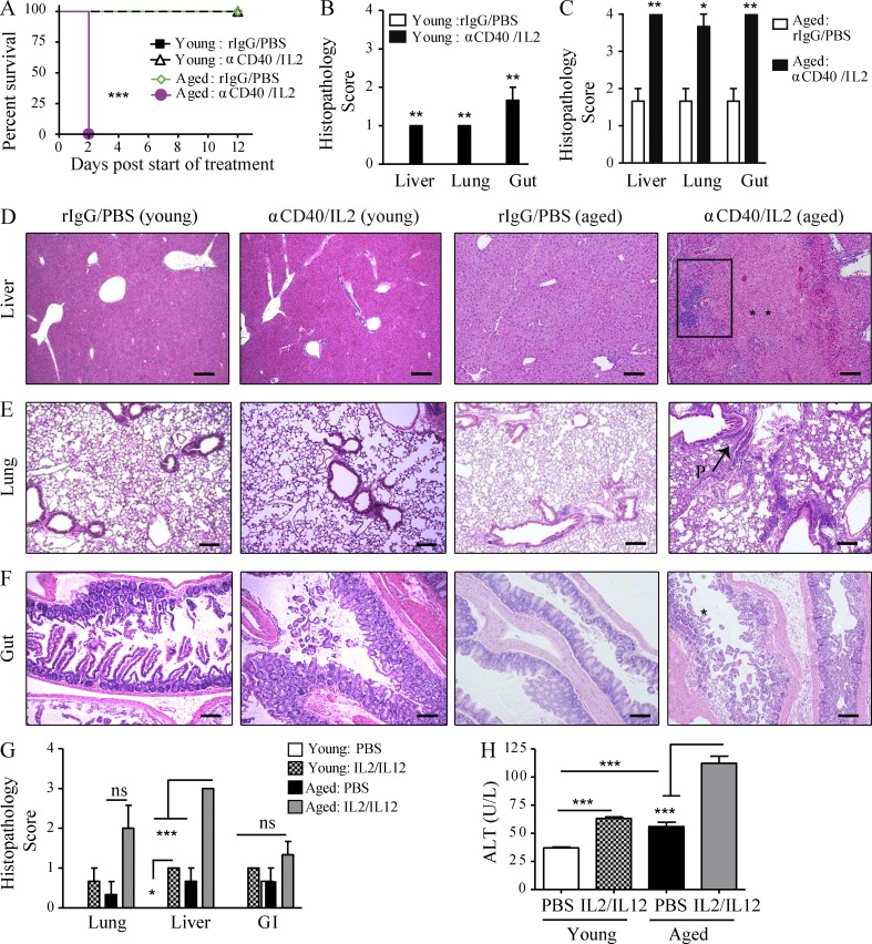
Increased mortality and multiorgan pathology in aged mice after high-dose anti-CD40/IL-2 treatment or after IL-2/IL-12. (A) Survival of young (4 mo) and aged (22 mo) naive C57BL/6 mice that received high-dose anti-CD40/IL-2 or rIgG/PBS, n = 5–8. (B–F) Three mice from each treated group in A were evaluated for histopathological differences in the liver at day 2. (B and C) Liver histopathology scoring (see Materials and methods) of young (B) and aged (C) mice. (D–F) Representative images of liver, lung, or gut H&E staining for young or aged mice. Bars, 500 µm. Liver necrosis (D) is indicated by ** and the rectangular area illustrating periportal lymphocytic aggregates. P: peribronchitis (E) in the lungs of aged mice. Gut mucosal erosion and damage (D) in aged mice is indicated by *. Data are representative of one of three independent experiments with similar results. Survival analysis was plotted according to the Kaplan-Meier method, and statistical differences were determined with the log-rank test. ***, P < 0.001; **, P < 0.01; *, P < 0.05. Ctrl: control. (G and H) Young (4 mo) and aged (17 mo) BALB/c mice received either PBS in the control group or 3 × 10⁵ IU IL-2 and 0.5 µg IL-12. (G) Histopathology of the lungs, livers, and gastrointestinal tracts collected at day 11 of treatment were scored and (H) serum ALT was quantified, n = 3. Values represent the mean ± SEM of one experiment. Bar graph (mean ± SEM) statistics were determined using two-way ANOVA with Bonferroni’s post-test, ***, P < 0.001; **, P < 0.01; *, P < 0.05. n.s.: not significant. &: P < 0.001 against all groups. A–F are representative of four and G and H of two independent experiments.

