Figure 3.
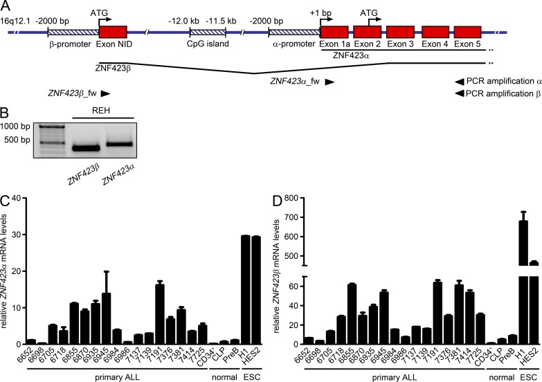
Identification of a novel ZNF423 isoform carrying a NID in ALL. (A) Structure of the ZNF423 locus. Red boxes, exons; striped blue boxes, regulatory region (promoter or CGI); arrows, position and orientation of primer oligonucleotides for PCR amplification of ZNF423 isoforms. Scheme is not drawn to scale. Adapted from UCSC genome browser. (B) PCR products of isoform-specific PCR with cDNA from REH cells. PCR products were separated by agarose gel electrophoresis and visualized by ethidium bromide staining. (C and D) Relative quantification of mRNA levels by qPCR using ZNF423 isoform-specific primers. ZNF423α and ZNF423β were normalized to B2M (2−ΔCt*1000). Error bars represent SD of technical triplicates.

