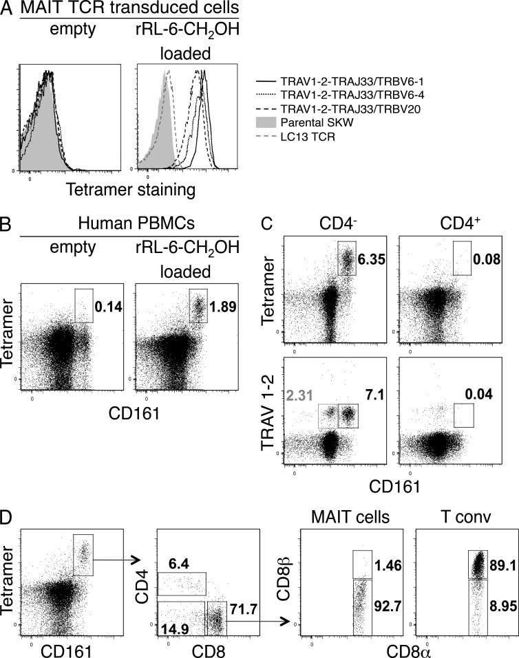
Figure 2.
Ag-specific identification of human MAIT cells. (A) Direct immunofluorescent staining of SKW3 cells expressing MAIT TCRs: TRAV1-2–TRAJ33 with TRBV6-1, TRBV6-4, or TRBV20, with empty (left) or rRL-6-CH2OH–loaded (right) human MR1 tetramer. Controls include nontransduced SKW3 (Parental SKW) and SKW3-transduced with an irrelevant TCR (LC13 TCR). (B) Human PBMCs were stained with CD3- and CD161-reactive mAbs and human MR1 tetramer. Shown are dot plots of CD3+ cells stained with empty (left) or rRL-6-CH2OH–loaded (right) human MR1 tetramer. Percentages of cells within boxed regions are indicated. Tetramer and CD161 staining are shown on the y and x axis, respectively. (C) Staining of human PBMCs comparing percentages of CD3+CD4− (left) and CD3+CD4+ (right) T cells detected by human MR1-Ag tetramer or TRAV1-2 mAb. Percentages of cells within (black) boxed regions are indicated. Also indicated is the percentage (gray) of CD3+CD4−TRAV1-2+CD161lo cells within the gray box (bottom left plot). Tetramer or TRAV1-2 and CD161 staining are shown on the y and x axis, respectively. Experiments in A–C were performed three times with similar results; shown are representative experiments. (D) Coreceptor expression on CD161+tetramer+ MAIT cells. CD3+CD161+tetramer+ cells were stained for cell surface expression of CD4, CD8α, and CD8β. CD8αα (CD8α+CD8β−) and CD8αβ (CD8α+CD8β+) expression on CD8+ MAIT cells was subsequently compared with expression on conventional CD8+ T cells (n = 6; shown is a representative staining experiment).

