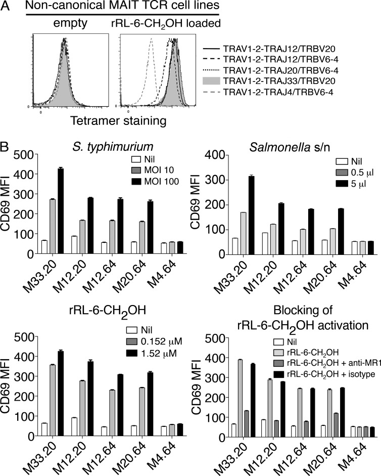
Figure 4.
T cells expressing TRAV1-2–TRAJ12 and TRAV1-2–TRAJ20 α-chains reconstitute features of canonical TRAV1-2–TRAJ33 MAIT cells. (A) Direct immunofluorescent staining of SKW3 cells expressing noncanonical MAIT TCRs: TRAV1-2–TRAJ12 with TRBV20 or TRBV6-4 and TRAV1-2–TRAJ20 with TRBV6-4, with empty (left) or human MR1-Ag tetramer (rRL-6-CH2OH loaded; right). Controls include a canonical MAIT TCR comprising TRAV1-2–TRAJ33 and TRBV20 α- and β-chains and a non-MAIT TCR comprising TRAV1-2–TRAJ4 and TRBV6-4 α- and β-chains. This experiment was performed twice with similar results. (B) SKW3 cells expressing noncanonical MAIT TCRs using TRAV1-2 joined with different TRAJs (M33.20, TRAV1-2–TRAJ33 plus TRBV20; M12.20, TRAV1-2–TRAJ12 plus TRBV20; M12.64, TRAV1-2–TRAJ12 plus TRBV6-4; M20.64, TRAV1-2–TRAJ20 plus TRBV6-4; and control non-MAIT TCR M4.64, TRAV1-2–TRAJ4 plus TRBV6-4) were activated by either infection with S. typhimurium (top left) or by addition of S. typhimurium supernatant (s/n; top right), rRL-6-CH2OH (bottom left), or rRL-6-CH2OH in the absence or presence of the anti-MR1 mAb (26.5) or an isotype control mAb (W6/32; bottom right). CD69 mean fluorescence intensity (MFI) is shown on the y axis. The tetramer-staining experiment was performed twice; the S. typhimurium infection, addition of S. typhimurium supernatant, and addition of rRL-6-CH2-OH experiments were performed once, three times, and three times, respectively, with similar results. Error bars show SEM of triplicates. Representative results are shown.

