
Figure 1. In the absence of IL-6, antigen-specific Foxp31 Treg cells expand at the expense of effector T cells (Teff cells)in vivo.
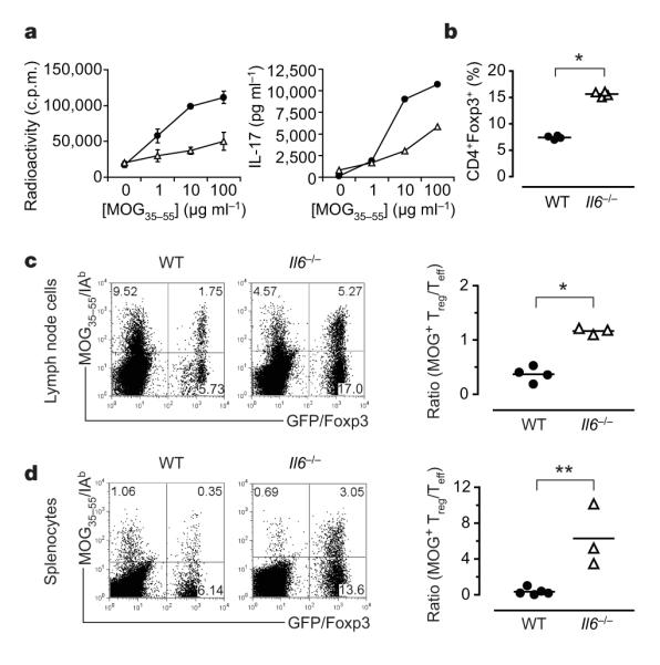
Foxp3gfp.KI (filled circles)or Il6−/− × Foxp3gfp.KI (Il6−/−; open triangles) mice were immunized with MOG35–55/CFA. a, Draining lymph-node cells were tested for MOG-specific proliferation and IL-17 production (means ± s.d. for triplicate determinations). b, The fraction of Foxp3/GFP+ T cells was determined ex vivo by flow cytometry (asterisk, P < 6 × 10⁻⁷; t-test). WT, wild type. c, d, Lymphnode cells (c) and splenocytes (d) from MOG35–55/CFA-immunized WT and Il6−/− mice were cultured for four days in the presence of MOG35–55 and stained with a MOG35–55/IAb tetramer. The ratios of antigen-specific Treg cells (MOG tetramer+CD4+Foxp3+) to Teff (MOG tetramer+CD4+Foxp3−) cells are presented (asterisk, P < 0.0003; two asterisks, P < 0.05; t-test).