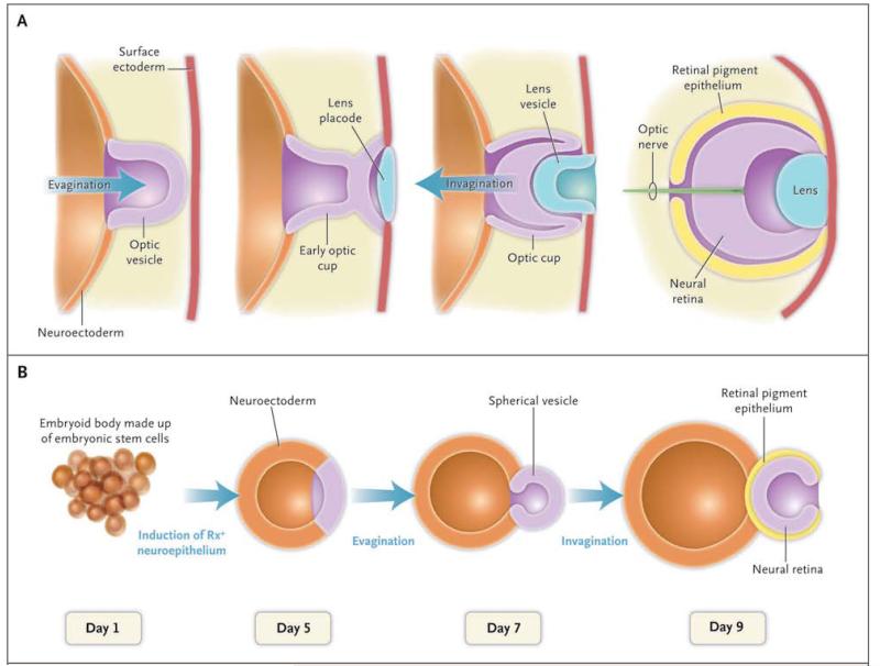
Figure 1. Formation of an Optic Cup in Vivo and in Vitro.
In Panel A, the embryonic neuroectoderm evaginates to form an optic vesicle. The interaction of the apical part of optic vesicle with the lens placode leads to the invagination and subsequent formation of an optic cup with a two-walled structure, consisting of retinal pigment epithelium on the outer wall and neural retina on the inner wall. In Panel B, over a period of 9 days, an optic-cup–like structure is formed in vitro in a three-dimensional culture of aggregates of embryonic stem cells. Self-aggregation of embryonic stem cells forms a spherical structure called an embryoid body. Subsequent invagination of its apical surface forms an optic cup with a two-walled structure consisting of retinal pigment epithelium and neural retina. The initial determination of eye primordium is defined by the expression of eye-specific transcription factor Rx.

