
Figure 4.
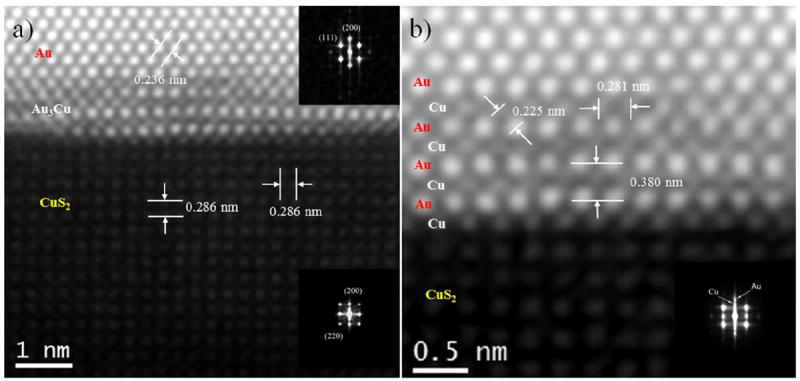
(a) Atomic-resolution HAADF-STEM image of core–shell nanoparticle different contrast exhibits Au-core, ordered Au3Cu interface region, and CuS2 surface layer, the corresponding inset fast Fourier transform (FFT) patterns exhibits the Au-core and CuS2 surface layer, (b) Close-up of the ordered Au3Cu alloyed shell region where the different contrasts of Au and Cu lattices are readily observable and inset shows the FFT of alloyed shell region.