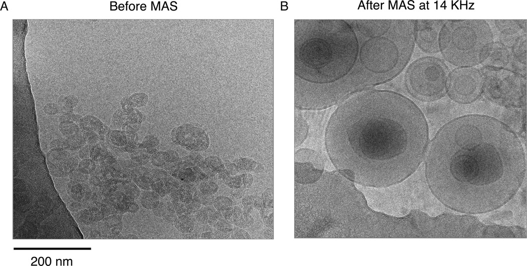
Figure 2.
Cryo-EM images of KcsA proteoliposomes in DOPE/DOPS lipid membranes are shown. In (A) an image of the sample before magic angle spinning shows predominantly small, unilamellar vesicles. Panel (B) shows an aliquot of the sample after magic angle spinning at 14 KHz for several days. The spinning causes these the vesicles to be converted into large multilamellar vesicles, but the bilayer remains intact.

