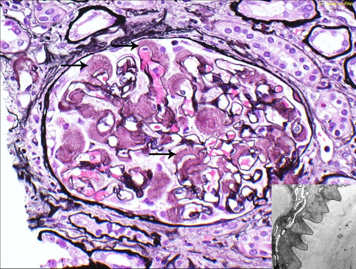
Figure 2.
Glomerular amyloid spicules. Glomerular amyloid spicules (arrows) result from parallel alignment of amyloid fibrils in the subepithelial zone perpendicular to the glomerular basement membrane. They were more common in AL/AH/AHL than other types of amyloidosis (Jones methenamine silver). The small image shows amyloid spicules by electron microscopy. AL/AH/AHL, light chain/heavy chain/heavy and light chain amyloidosis. Original magnification, ×400 in image; ×23,000 in inset.

