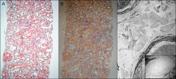
Figure 3.
ALECT2. (A) This case of ALECT2 exhibits diffuse cortical interstitial amyloid deposition. The glomerulus on the left is also globally involved (Congo red stain). (B) The Congo red-positive amyloid deposits show apple-green birefringence when viewed under polarized light. (C) Electron microscopy shows several interstitial aggregates of amyloid deposits. The small image is a higher magnification showing the fibrillar nature of deposits. ALECT2, leukocyte chemotactic factor 2 amyloidosis. Original magnification, ×100 in A and B; ×5800 in C; ×46,000 in C inset.

