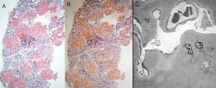
Figure 5.
AFib. (A) There is diffuse obliterative glomerular involvement by amyloidosis in this case of AFib (Congo red stain). (B) The Congo red-stained sections from the case shown in A exhibit apple-green birefringence when viewed under polarized light. (C) Electron microscopy shows marked mesangial and glomerular capillary wall amyloid deposition. Despite the subtotal obliteration of glomerular capillary lumina by amyloid deposits, no amyloid spicules are seen. AFib, fibrinogen A α chain amyloidosis. Original magnification, ×100 in A and B; ×1900 in C.

